Magnetic resonance imaging (MRI) didn’t start as the technology we now commonly know. Researchers Felix Bloch and Edward Purcell first discovered the magnetic resonance phenomena in 1946 and later harnessed the abilities of magnetic resonance to analyze chemicals, leading to their Nobel Prize in 1952. Later on, scientists discovered the same technique could be used to visualize different human tissues. By 1973, aided by the rapid technological progression of computers, researchers developed the MRI that we now use today. Then, in 2003, the MRI led to another Nobel Prize, this time awarded to researchers Paul Lauterbur and Peter Mansfield for developing MRI as a diagnostic tool.
What is an MRI?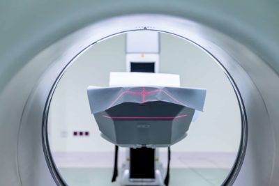
An MRI uses a strong magnetic field and directs the field at a specific area of the object or person of interest. As the magnetic field enters different tissues and fluids within the body, hydrogen atoms become excited similarly to how a smaller magnet becomes excited as a larger magnet inches closer. Depending on the tissue or fluid that the hydrogen atoms are in, the atoms return to a resting state at different rates as the magnetic field is turned on and off several times. This allows a computer to analyze the different rates and create an image that shows a contrast in the different tissues and fluids.
The process of turning a magnetic field on and off is repeated numerous times over each body part being imaged and is then completed in multiple planes of motion. Collecting images through each plane of motion allows a doctor the ability to see an injury, or lack thereof, from each viewpoint. Resultantly, a doctor would then be able to develop the most accurate diagnosis to create the most effective treatment protocol.
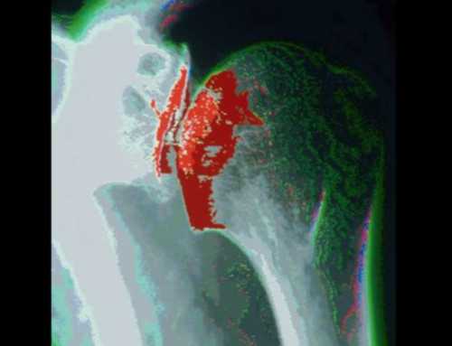
MRI’s differ from x-rays in that they are more effective at demonstrating abnormalities in tissue samples rather than bone samples. For example, a torn or degenerative meniscus may present a decreased joint space in the knee as apparent on x-ray, but an MRI would show the actual torn or missing tissue of the meniscus that causes the decreased joint space. Without the MRI the diagnosis would not be confirmed and an effective treatment protocol could not begin.
How does an MRI show an injury?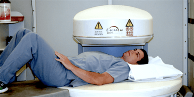
Trained health professionals can locate a severed or partially torn muscle, tendon (connects muscle to bone), or ligament (connects bone to bone) by comparing the shape of the tissue of interest to that of a normal, healthy tissue. For example, a torn anterior cruciate ligament (ACL) in the knee may appear frayed at one end if torn. The frayed image appears as fluid from the knee flows around the torn ACL to encompass the tissue and project different rates of hydrogen atoms returning to a resting state as explained above, leaving only the tissue to present a darker contrasting appearance.
Open vs. Closed MRI’s
There are two kinds of MRI systems, open and closed, which differ by just a few key points.
- Comfort: As one might expect from the name, an open MRI offers a much more comfort. For someone who is injured, sick, or claustrophobic, an open MRI presents a greater amount of restfulness when compared to the cramped spacing of a closed MRI system. As a result, a patient who is able to remain calmer during MRI imaging often gets a better picture of the area of interest due to there being less movement during the process. In addition, the open space allows imaging technicians to place patients into positions that improve the quality of images, something that can’t always be done in a closed MRI.
- Imaging Power: On the other side, closed MRI’s have greater imaging power (1.5 Tesla in closed MRI compared to 0.3 Tesla in open). Larger imaging power can result in a clearer MRI image if scanning a deep tissue sample. However, in the case of orthopedics, an open MRI’s power of around 0.3 Tesla is sufficient to obtain clear images of bones, muscles, and ligaments.
- Cost: A sometimes significant cost difference is found between open and closed MRI’s due to the larger magnets closed MRI’s. Less upkeep is needed to maintain an open MRI system and can result in a price decrease of 40-50% when compared to the cost of a closed system.
Washington Orthopaedic Center offers an on-site open MRI machine in Centralia to improve convenience and comfort. Our imaging technologists are even kind enough to play your music of choice during your MRI to improve your experience, as the process typically takes between 30 – 60 minutes. Washington Orthopaedic Center also contains an x-ray imaging center to accompany the MRI machine. For the greatest convenience, without the hassle of wandering through a hospital, turn to Washington Orthopaedic Center for your care.

