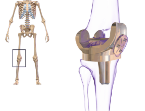
Runner’s knee or patellofemoral pain syndrome is a common knee problem found in many athletes, especially new runners. Patellofemoral pain is seen most commonly in athletes and runners; this constitutes 25% of all identified knee injuries [1]. PFP affects women more than mend in a 2:1 ratio and is seen most commonly in adolescents [2].
What causes patellofemoral pain syndrome?
Patellofemoral pain syndrome is due to the kneecap (patella) not tracking properly in the femoral trochlea, causing rubbing of the cartilage in abnormal ways. The cartilage on the underside of the kneecap is there to let your knee cap glide back in forth between the groove on the femur. The abnormal tracking of the kneecap is usually due to a muscle imbalance of the Quadricep muscles pulling the knee cap usually to the outside of the knee. Can also be caused by tight hamstrings, poor foot mechanics, hip problems or various other causes.
There is also various ligaments that attach to the inside and outside of the patella, which can be injured, stretched, or torn in trauma which can lead to kneecap instability. These are best assessed with an MRI.
Symptoms
Most common symptoms of patellofemoral pain syndrome include a dull or achy type pain to the anterior or front of the knee. The pain is usually worse with squatting movements, stairs, and walking up or down hills. You may experience knee swelling.
What a practitioner may observe during your appointment
A clinician may observe the way you walk, looking for any abnormalities in foot, knee, or hip mechanics. You will also assessed for evidence of weakness to the quad muscles and reduction in the range of motion of your knee. A practitioner will assess the knee ligament for instability. Address any possible meniscus issues causing referred pain to the knee cap. Also assess for any kneecap instability or patella pain.
Imaging:
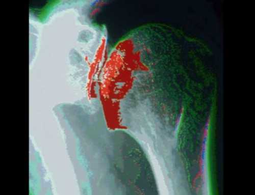
Possible imaging that may be obtained to assess patellofemoral pain include an x-ray to look at knee cap alignment, arthritis, or possible fractures that may be seen on x-ray. Occasionally an MRI or bone scan will be obtained to look at the cartilage or soft tissues and ligaments. These are usually only obtained if patient is not progressing with treatment.
Treatment:
There are many treatment options for this problem. First and foremost is rest. If you knee is acutely in pain, you should rest and ice it. Your practitioner may prescribe some anti-inflammatory type medications to help reduce some of the inflammation that is occurring in your knee. One of the best treatments for this issue is working with a physical therapist to help and strengthen the quad muscles to realign the patella into the femoral groove. Other treatments include bracing, additional treatments to the hips or feet. If you are not getting better after some of these modalities, an MRI may be obtained to assess the ligaments. Occasionally surgery may be indicated.

Lukas Steffan PA-C
If you or a friend may think you are having issues with patellofemoral pain, you can schedule with one of our providers for evaluation or treatment at Washington Orthopedic Center! Contact us now to set up an appointment at 360-736-2889
Article by Lukas Steffan (PA-C)
References
[1] Baguie, & Brukner. (1997). Injuries presenting to an Australian sports medicine centre: a 12-month study. Retrieved from https://www.ncbi.nlm.nih.gov/pubmed?term=9117522
[2] DeHaven KE, & Lintner DM. (1986). Athletic injuries: comparison by age, sport, and gender. [Abstract]. Retrieved February 21, 2018.


